When a doctor examines a tissue sample under a microscope, they are looking for clues. These clues, hidden at a cellular level, tell a story about what’s happening inside the body. One of the most significant clues they can find is a specific type of tissue damage called fibrinoid necrosis. While the name sounds complicated, understanding what it is and what causes it can shed light on several serious medical conditions. It’s a signpost that points clinicians toward a correct diagnosis and an effective treatment plan.
This type of tissue damage is not a disease in itself, but rather a finding a piece of evidence left behind by an underlying problem. It signals that a destructive process is underway, most often within the walls of blood vessels. Recognizing fibrinoid necrosis is a critical step for pathologists, as it dramatically narrows down the list of potential causes, allowing for more focused care for the patient.
What Exactly Is Fibrinoid Necrosis?
To understand fibrinoid necrosis, let’s first break down the term. “Necrosis” is the medical word for the death of body tissue. It’s an irreversible injury, and the cells can’t be repaired. The word “fibrinoid” means “fibrin like.” Fibrin is a protein that your body uses to form blood clots and stop bleeding. When a pathologist sees fibrinoid necrosis, they see an area of tissue that has died and is now filled with a substance that looks bright pink, glassy, and similar to fibrin under the microscope.
However, this substance isn’t just fibrin. It’s actually a complex mixture. Imagine a small blood vessel as a tiny garden hose. If this hose gets damaged, its walls begin to leak. In the case of fibrinoid necrosis, the vessel wall is so damaged that large molecules from the blood plasma like antibodies, complement proteins, and yes, fibrin leak out and get deposited into the vessel wall itself. This collection of proteins and cellular debris creates the characteristic “fibrin like” appearance.
This process completely disrupts the normal structure of the tissue. The cells die, and the organized layers of the blood vessel wall are replaced by this amorphous, pink material. This isn’t just a cosmetic change, it severely weakens the blood vessel, making it prone to rupture, and it can block the flow of blood to vital organs.
A Look Under the Microscope: What Does It Look Like?
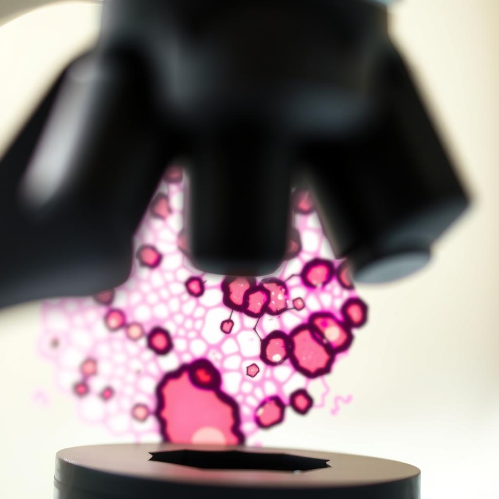
For a pathologist, identifying fibrinoid necrosis is a visual skill. When a tissue sample (a biopsy) is prepared for examination, it’s sliced incredibly thin, placed on a glass slide, and stained with special dyes to make different components visible. The most common stain is called Hematoxylin and Eosin (H&E).
- Hematoxylin stains cell nuclei a purplish blue.
- Eosin stains the cytoplasm and extracellular matrix (like collagen) various shades of pink.
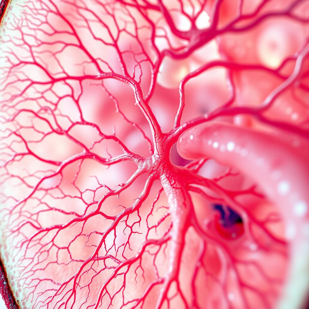
In a healthy blood vessel, you would see distinct layers with neatly arranged cell nuclei. Under an H&E stain, areas of fibrinoid necrosis stand out dramatically. They appear as a bright, intensely eosinophilic (pink) band or smudge within the vessel wall. All the fine details are lost. The normal blue staining nuclei of the smooth muscle cells disappear, replaced by this structureless, protein heavy deposit. It’s a clear visual signal that severe, acute injury has occurred.
The Core Causes: Why Does Fibrinoid Necrosis Happen?
The development of fibrinoid necrosis primarily boils down to two major pathways: severe immune reactions or extreme physical damage to blood vessels. Both pathways lead to a final common outcome of plasma proteins leaking into and destroying the vessel wall.
Immunologic Damage: When the Body Attacks Itself
The most common cause of fibrinoid necrosis is an immune system malfunction. Specifically, it’s often linked to what is known as a Type III hypersensitivity reaction. Think of it this way: your immune system creates antibodies to fight off invaders like bacteria and viruses. These antibodies are like custom made handcuffs for a specific bad guy (the antigen). Normally, this works perfectly.
In some autoimmune diseases, the body mistakenly creates antibodies against its own proteins. These antibodies and self antigens link up to form small clumps called immune complexes. When these complexes are formed in large amounts, they can circulate in the blood and get stuck in the walls of small blood vessels. Once lodged, these complexes trigger a powerful inflammatory reaction, attracting destructive white blood cells to the site and leading to the severe vessel damage that results in fibrinoid necrosis.
Several conditions are classic examples of this process:
Systemic Lupus Erythematosus (SLE)
Lupus is a chronic autoimmune disease where the immune system attacks multiple organs, including the skin, joints, kidneys, and blood vessels. The deposition of immune complexes in the small vessels of the kidneys is a hallmark of lupus nephritis, and finding fibrinoid necrosis in a kidney biopsy is a sign of severe, active disease.
Polyarteritis Nodosa (PAN)
PAN is a form of vasculitis, which means inflammation of the blood vessels. It typically affects medium sized arteries. The inflammation is so intense that it causes segmental fibrinoid necrosis throughout the vessel wall. This weakens the artery, leading to bulges (aneurysms) and potential rupture. Seeing this type of necrosis is a key feature used to diagnose PAN.
Rheumatic Fever
This condition can develop after a streptococcal throat infection. The immune system, in its effort to fight the bacteria, accidentally cross reacts with proteins in the heart tissue. This leads to the formation of characteristic inflammatory lesions in the heart called Aschoff bodies, which often have a central zone of fibrinoid necrosis.
Rheumatoid Arthritis
In some people with severe rheumatoid arthritis, firm lumps called rheumatoid nodules can form under the skin, often over joints like the elbow. These nodules characteristically have a central area of fibrinoid necrosis surrounded by inflammatory cells.
Severe Vascular Injury: The Pressure Problem
The second major cause of fibrinoid necrosis is not related to the immune system but to sheer physical force, specifically extremely high blood pressure. This is a non immunologic pathway where mechanical stress causes the damage.
Malignant Hypertension
This is a medical emergency where blood pressure rises to dangerously high levels very quickly. Imagine turning the pressure in a firehose up so high that the lining of the hose itself starts to fray and tear. That’s analogous to what happens in the body’s smallest arteries (arterioles), particularly in the kidneys. The extreme pressure directly injures the vessel walls, forcing plasma proteins into them and causing fibrinoid necrosis. This is a tell tale sign of hypertensive crisis.
Pre eclampsia and Eclampsia
These are serious blood pressure disorders that can occur during pregnancy. The high blood pressure and other factors can cause injury to the blood vessels of the placenta. Pathologists often find fibrinoid necrosis in these placental vessels, which reflects the vascular damage that can impair blood flow to the developing fetus.
Here is a table summarizing the main causes:
| Causative Pathway | Mechanism | Associated Conditions |
|---|---|---|
| Immunologic | Immune complex deposition (Type III Hypersensitivity) | Systemic Lupus Erythematosus (SLE), Polyarteritis Nodosa (PAN), Rheumatic Fever, Rheumatoid Arthritis |
| Non Immunologic (Vascular Injury) | Extreme mechanical stress from high blood pressure | Malignant Hypertension, Pre eclampsia/Eclampsia |
The Significance of Finding Fibrinoid Necrosis
Finding fibrinoid necrosis in a biopsy is a very important event. It’s not a subtle finding, it’s a clear indicator of a severe and active disease process. For a clinician, receiving a pathology report that mentions this finding is a major red flag.
First, it immediately narrows the diagnostic possibilities. For instance, if a patient has kidney failure and a biopsy shows fibrinoid necrosis, the doctor will strongly suspect conditions like severe lupus nephritis, a vasculitis like PAN, or malignant hypertension. It helps distinguish these aggressive conditions from other, more slowly progressing forms of kidney disease.
Second, it indicates the severity of the condition. The presence of fibrinoid necrosis implies that the tissue damage is acute and significant. This often prompts more aggressive treatment to get the inflammation or blood pressure under control quickly to prevent further organ damage. It signals that the disease is not in a quiet or smoldering phase but is actively causing destruction.
How Is the Underlying Condition Treated?
It’s crucial to remember that you don’t treat the fibrinoid necrosis itself, you treat the disease that is causing it. The necrosis is the result of the battle, not the battle itself. Once the underlying cause is addressed, the body can begin the healing process, which usually involves clearing away the dead tissue and replacing it with scar tissue.
The treatment strategies vary widely depending on the cause:
- For Autoimmune Conditions (like SLE or PAN): The goal is to suppress the overactive immune system. This is typically done with powerful anti inflammatory and immunosuppressive drugs. Corticosteroids (like prednisone) are often used initially to quickly reduce inflammation, followed by other medications like cyclophosphamide or rituximab to achieve long term control.
- For Malignant Hypertension: This is a medical emergency that requires immediate hospitalization. The goal is to lower the blood pressure safely and quickly using intravenous antihypertensive medications. The patient will then need to be on long term oral medications to manage their blood pressure.
- For Pre eclampsia/Eclampsia: The most definitive treatment is the delivery of the baby and placenta. Medications to lower blood pressure and prevent seizures may also be used to manage the mother’s condition until delivery is possible.
Frequently Asked Questions (FAQ)
Is fibrinoid necrosis a type of cancer?
No, fibrinoid necrosis is not cancer. It is a form of cell death and tissue damage caused by inflammation or severe injury, whereas cancer is characterized by uncontrolled cell growth. While it is a very serious finding, it is not related to malignancy.
Is fibrinoid necrosis reversible?
The necrosis itself the cell death is irreversible. The dead cells cannot be brought back to life. However, the process that causes fibrinoid necrosis can be stopped. By treating the underlying autoimmune disease or controlling the severe hypertension, further damage can be prevented. The body will then heal the damaged areas, typically by forming scar tissue.
Where is fibrinoid necrosis most commonly found?
Fibrinoid necrosis is almost always found in the walls of blood vessels, particularly small arteries and arterioles. This is because these are the primary sites where immune complexes get deposited and where the effects of severe hypertension are most pronounced. It can be seen in virtually any organ, but it is particularly common in the kidneys, heart, skin, and placenta, depending on the specific disease.
Can you have fibrinoid necrosis without an autoimmune disease?
Yes. The classic non immune cause is malignant hypertension. In this situation, the cause is extreme mechanical stress on the blood vessel walls, not an attack from the immune system. This shows that while frequently linked to autoimmunity, fibrinoid necrosis is ultimately a pattern of vascular injury with more than one possible cause.
Conclusion
Fibrinoid necrosis is a specific and serious pattern of tissue death that provides vital information to doctors. Seen as a bright pink, smudgy deposit under the microscope, it represents severe damage to blood vessel walls. This damage is most often triggered by one of two things: a powerful attack from the body’s own immune system in conditions like lupus and vasculitis, or extreme physical stress from dangerously high blood pressure. While fibrinoid necrosis itself is a sign of irreversible damage, its discovery is a critical diagnostic clue. It alerts clinicians to the presence of a severe, active disease, prompting them to begin the aggressive treatment necessary to control the underlying condition and prevent further harm to the body’s organs.

Timo is the founder of LiteDietPlan.com, where smart nutrition meets simple living.