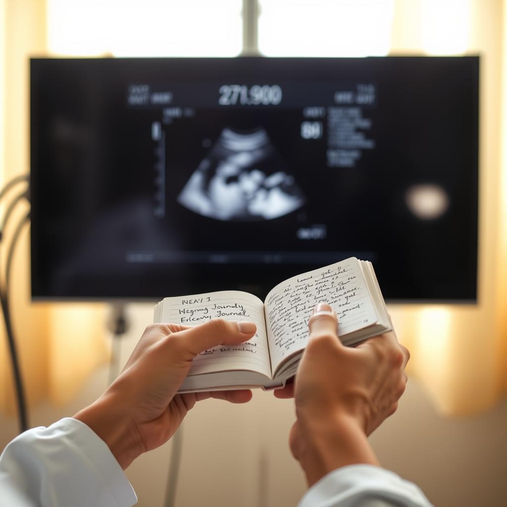
Hearing a baby’s heartbeat for the first time is a profound moment in any pregnancy journey. It’s a rhythmic, reassuring sound that makes everything feel more real. While many are familiar with the electronic whooshing sound from a Doppler machine, there is another tool that has been used for over a century to connect with this new life: the fetoscope. This simple looking device comes in two dramatically different forms, one a classic tool of midwifery and the other a high tech instrument that makes modern medical miracles possible. Understanding the fetoscope reveals a fascinating story of medical innovation.
The Tale of Two Fetoscopes: From Simple Horn to High Tech Tool
When you hear the word “fetoscope,” it’s important to know which one is being discussed. The term can refer to two completely different medical instruments that serve very distinct purposes. One is a non electric, acoustic device for listening, while the other is an advanced, fiber optic camera used for surgery inside the womb. Confusing the two would be like mistaking a pair of binoculars for the Hubble Space Telescope, both let you see things far away, but their capabilities are worlds apart.
The Acoustic Fetoscope: Listening to Life’s First Rhythms
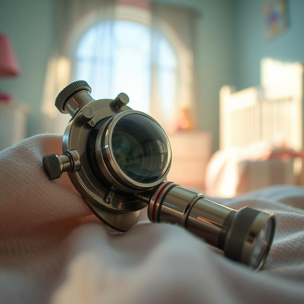
The original fetoscope is an acoustic device, meaning it works by simply capturing and amplifying sound waves. It requires no batteries, electricity, or complex electronics. Think of it as a specialized type of stethoscope. The most recognizable version is the Pinard horn, which looks like a simple wooden or metal cone. Another common type is the DeLee Hillis fetoscope, which looks more like a traditional stethoscope but has a weighted metal band that goes on the listener’s head to improve sound conduction.
This type of fetoscope is still favored by many midwives and some doctors. They appreciate its simplicity and the fact that it doesn’t use ultrasound waves. Using an acoustic fetoscope is a skill that requires practice, as the practitioner must find the precise spot on the abdomen to clearly hear the heartbeat. This hands on approach also helps them determine the baby’s position within the womb.
The Modern Endoscopic Fetoscope: A Window into the Womb
The second, more modern device is the endoscopic fetoscope. This is a highly sophisticated surgical instrument. It consists of a very thin, tube like instrument called an endoscope that contains a fiber optic light source, a tiny camera, and small channels to pass surgical tools through. This advanced fetoscope is not used for routine check ups but for performing minimally invasive surgery on a fetus while it is still in the uterus.
During a procedure called a fetoscopy, this fetoscope is inserted through a tiny incision in the mother’s abdomen and uterus. The camera transmits a live video feed to a monitor, giving surgeons a direct, magnified view inside the amniotic sac. This allows them to diagnose and treat complex medical conditions before the baby is even born, offering hope for situations that were once untreatable.
How Is an Acoustic Fetoscope Used?
Using a traditional acoustic fetoscope is both an art and a science. A midwife or doctor will first palpate, or feel, the mother’s abdomen to get a sense of the baby’s position. They are trying to locate the baby’s back, as this is the best place to pick up the sound of the heart. Once they have an idea of the location, they place the wide end of the fetoscope firmly on the abdomen.
The practitioner then presses their ear to the other end and listens intently, moving the fetoscope slightly until the heartbeat is found. It takes a trained ear to distinguish the rapid “lub dub” of the fetal heart from other sounds, like the mother’s heartbeat or digestive noises. Besides just confirming the heart rate, this method can help the practitioner:
- Confirm the baby’s position (e.g., head down or breech).
- Identify if there are multiple babies.
- Listen for signs of fetal distress during labor.
Fetoscope vs. Doppler: What’s the Difference?
The most common tool used to listen to a fetal heartbeat today is the handheld Doppler device. It works very differently from an acoustic fetoscope and each has its own set of advantages and limitations. A Doppler transmits high frequency sound waves (ultrasound) into the body, which bounce off moving objects, like the baby’s heart valves and blood cells. The device detects the change in frequency of the returning waves and converts it into an audible sound.
Here is a breakdown of the key differences:
| Feature | Acoustic Fetoscope | Handheld Doppler |
|---|---|---|
| How it Works | Passive sound amplification | Ultrasound waves (Doppler effect) |
| Power Source | None | Batteries |
| Safety | No energy waves, no known risks | Generally considered safe, but uses ultrasound energy |
| When It Can Be Used | Around 18 20 weeks of gestation | As early as 8 12 weeks of gestation |
| What You Hear | The actual “lub dub” sound of the heart valves closing | An amplified, electronic sound of blood movement |
| Additional Information | Helps determine the baby’s position | Primarily used for heart rate confirmation |
Understanding Fetoscopy: The Surgical Use of a Modern Fetoscope
While the acoustic fetoscope offers a gentle way to monitor pregnancy, the endoscopic fetoscope is at the forefront of medical intervention. The procedure, known as fetoscopy, is a form of fetal surgery. It is reserved for treating severe and often life threatening birth defects that could cause irreversible damage if left untreated until after birth. This is a highly specialized field of medicine performed at only a few top medical centers around the world.
The goal of using a surgical fetoscope is to correct a problem with the least amount of disruption to the pregnancy. By making a very small incision, surgeons can avoid the risks associated with traditional open fetal surgery, where the uterus is opened more widely. The use of a fetoscope has revolutionized the treatment of several serious conditions.
What Conditions Are Treated with a Fetoscope?
Fetal surgeons use a fetoscope to perform delicate procedures on a baby smaller than the palm of their hand. The video guidance allows for incredible precision in a space that was previously inaccessible without major surgery. Some of the groundbreaking procedures performed with a fetoscope include:
- Twin to Twin Transfusion Syndrome (TTTS): In some identical twin pregnancies, blood vessels in the shared placenta are connected unevenly, causing one twin to get too much blood and the other too little. Surgeons can use a laser passed through the fetoscope to seal off these connecting vessels, correcting the imbalance and saving both babies.
- Congenital Diaphragmatic Hernia (CDH): This is a condition where a hole in the diaphragm allows abdominal organs to move into the chest, preventing the lungs from growing properly. Using a fetoscope, surgeons can perform a procedure called FETO (Fetoscopic Endoluminal Tracheal Occlusion), where a tiny balloon is temporarily placed in the fetus’s trachea. This encourages lung growth before birth.
- Spina Bifida: In some cases of this condition where the spinal cord doesn’t close properly, a fetoscope can be used to guide surgeons as they place a patch over the opening on the baby’s back. This can reduce nerve damage and improve outcomes after birth.
What to Expect During a Fetoscopy Procedure
A fetoscopy is a major medical procedure that requires a highly skilled team. The pregnant person is typically given regional (epidural) or general anesthesia. The surgeons use ultrasound continuously to monitor the fetus and guide their movements. They make a tiny incision, often just a few millimeters wide, on the abdomen and insert a thin tube called a cannula. The fetoscope is then passed through the cannula and into the amniotic sac.
While looking at the monitor, the surgeons can introduce laser fibers, graspers, or other miniature instruments through the channels in the fetoscope to perform the necessary repair. After the procedure is complete, the instruments are removed, and the small incision is closed. The patient is then monitored very closely in the hospital for several days to watch for signs of complications like preterm labor.
Risks and Benefits of Using a Fetoscope
The risks and benefits associated with a fetoscope depend entirely on which type is being used. For the acoustic device, the profile is one of safety and simplicity, while for the surgical device, it involves a complex balance of life saving potential against serious risks.
Acoustic Fetoscope: Simplicity and Safety
The primary benefit of the traditional fetoscope is its complete safety. It emits no energy and simply funnels sound, so there are no risks to the parent or baby. It is inexpensive, durable, and provides a direct connection to the sound of the baby’s heart. For many, hearing the organic “lub dub” sound is a more natural bonding experience. The main limitation is that it can’t detect the heartbeat until later in the pregnancy and requires a skilled user to be effective.
Endoscopic Fetoscopy: A High Reward, High Risk Procedure
For fetoscopy, the benefits can be immense. The procedure can save the lives of fetuses with conditions that would otherwise be fatal. By intervening before birth, it can prevent or reduce severe organ damage and dramatically improve a child’s quality of life. However, it is an invasive procedure with significant risks. These include premature rupture of the amniotic sac membranes (PPROM), preterm labor, infection, and, in some cases, the loss of the pregnancy. These risks are weighed carefully by the medical team and the family before proceeding.
Frequently Asked Questions (FAQ)
When can you first hear a baby’s heartbeat with a fetoscope?
An acoustic fetoscope can typically pick up the fetal heartbeat between 18 and 20 weeks of pregnancy. This is later than a Doppler, which can often detect the heartbeat as early as 8 to 12 weeks.
Is a fetoscope the same as a stethoscope?
They are similar in that both are acoustic devices used for listening to internal body sounds. However, a fetoscope is specifically designed to better capture the higher frequency, lower volume sounds of a fetal heart through the abdomen. The DeLee Hillis fetoscope even includes a headpiece to maximize sound conduction.
Does using a fetoscope hurt?
Using an acoustic fetoscope is completely painless. The practitioner will press it firmly against your abdomen, but it should not cause any discomfort. The surgical procedure of fetoscopy, however, involves an incision and is performed with anesthesia to manage pain.
Who performs a surgical fetoscopy?
This is a highly specialized procedure performed by a team led by a maternal fetal medicine specialist or a pediatric surgeon with advanced training in fetal surgery. These procedures are only done at major hospitals with dedicated fetal treatment centers.
Why would my midwife use a fetoscope instead of a Doppler?
A midwife might choose a fetoscope for several reasons. Some prefer to avoid exposing the fetus to ultrasound waves, even though Dopplers are considered safe. Others appreciate that using a fetoscope requires hands on skills that also help them determine the baby’s position and lie, providing more information than just a heart rate. It also fosters a different kind of connection during prenatal appointments.
Conclusion
The fetoscope is a remarkable example of how a single term can encompass both the roots of medicine and its most advanced frontiers. On one hand, the acoustic fetoscope remains a simple, elegant tool that allows a provider to listen to the fundamental rhythm of life without any electricity or waves, fostering a direct connection between caregiver and baby. On the other hand, the modern endoscopic fetoscope is a technological marvel a tiny camera and surgical port that allows doctors to perform life saving operations before a baby takes its first breath. Whether used to simply listen or to surgically intervene, the fetoscope plays a vital role in prenatal care, offering reassurance, information, and hope to families.