
Deep within the human abdomen lies a complex network of blood vessels, working silently to power our core muscles. Among these, the inferior epigastric artery plays a starring role, acting as a critical lifeline for the anterior abdominal wall. Understanding its path, its job, and its importance in medicine reveals just how interconnected our anatomy is, where a single artery can be the difference between a successful surgery and a serious complication.
What Exactly is the Inferior Epigastric Artery?
Think of your body’s circulatory system as a massive, intricate highway system. The aorta is the main interstate, and branching off it are major state highways, which in turn lead to smaller county roads. In this analogy, the inferior epigastric artery is a vital county road that supplies a specific, important neighborhood: the lower front part of your abdominal wall, including the “six pack” muscles.
This artery is part of a pair, with one on the right side and one on the left. It’s not a massive vessel, but its location makes it one of the most clinically significant arteries in the abdomen. Surgeons, radiologists, and anatomists pay close attention to the inferior epigastric artery because of its role as a key landmark and its potential for injury during medical procedures.
Mapping the Journey: Location and Path of the Inferior Epigastric Artery
To truly understand this artery, you need to follow its path. Its journey is relatively short but passes through several critical anatomical structures. Knowing its origin and course is fundamental for any medical professional working in the abdominal region.
Origin: Where It All Begins
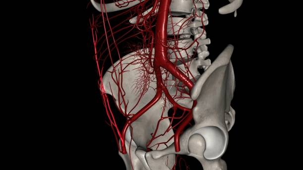
The inferior epigastric artery begins its journey deep in the pelvis. It branches off a much larger artery called the external iliac artery. This happens just before the external iliac artery passes under a tough band of tissue called the inguinal ligament to become the femoral artery, which is the main blood supply to the leg.
This starting point is significant because it’s in a busy area, surrounded by nerves, veins, and lymphatic structures. The artery is accompanied by the inferior epigastric vein, which runs alongside it, carrying deoxygenated blood back toward the heart.
The Course: A Path Up the Abdominal Wall
After branching off, the inferior epigastric artery curves forward and then travels upward. Its primary destination is the back surface of the rectus abdominis muscle the long, flat muscle that runs vertically down the front of your abdomen.
As it ascends, it pierces a layer of connective tissue called the transversalis fascia. It continues its climb within a protective sleeve known as the rectus sheath, nestled between the muscle and the posterior layer of the sheath. Finally, high up in the abdomen near the belly button, the inferior epigastric artery ends by forming connections (anastomoses) with the superior epigastric artery, which comes down from the chest. This connection creates a continuous blood supply along the entire front of the abdomen.
Key Anatomical Facts of the Inferior Epigastric Artery
| Feature | Description |
|---|---|
| Origin | External iliac artery |
| Main Function | Supplies blood to the lower anterior abdominal wall |
| Primary Muscle Supplied | Rectus abdominis muscle |
| Accompanies | Inferior epigastric vein |
| Termination | Anastomoses with the superior epigastric artery within the rectus sheath |
The Branches: Supplying the Abdominal Wall
Like a river feeding smaller streams, the inferior epigastric artery gives off several smaller branches along its path to nourish surrounding tissues. These branches are essential for the health of the abdominal wall.
- Cremasteric Artery: In males, this small branch travels with the spermatic cord to supply the cremaster muscle and other coverings of the cord. In females, a similar but smaller artery supplies the round ligament of the uterus.
- Pubic Branch: This branch runs down towards the pubic bone, supplying it with blood. It also forms a connection with the pubic branch of another artery, the obturator artery.
- Muscular Branches: Numerous small branches peel off to supply the rectus abdominis muscle and the surrounding flat muscles of the abdominal wall (the obliques and transversus abdominis).
- Cutaneous Branches: These branches pierce through the muscle and supply the overlying skin of the lower abdomen.
What Does the Inferior Epigastric Artery Do? Key Functions
The primary function of the inferior epigastric artery is straightforward: it delivers oxygen rich blood to the structures of the lower anterior abdominal wall. Without this constant supply, the muscles would weaken, and the skin and tissues would not be able to repair themselves.
Its main jobs include:
- Nourishing the Rectus Abdominis: This is its most important task. The rectus abdominis muscle is crucial for posture, flexing the torso, and protecting the internal organs.
- Supplying Abdominal Skin: It keeps the skin over your lower belly healthy and alive.
- Supporting Peritoneum: It provides blood to the peritoneum, the thin membrane that lines the inside of the abdominal cavity.
The anastomosis, or connection, with the superior epigastric artery is also a critical feature. It creates a backup route for blood flow. If one artery were partially blocked, this connection could help blood from the other artery reach the affected area, preventing tissue damage.
Clinical Significance: Why This Artery Matters in Medicine
While fascinating from an anatomical perspective, the true importance of the inferior epigastric artery becomes clear in a clinical setting. Its predictable location makes it both a helpful landmark and a potential hazard during medical procedures.
Inguinal Hernias and the Hesselbach’s Triangle
An inguinal hernia is a bulge that occurs when tissue, such as part of the intestine, protrudes through a weak spot in the abdominal muscles. The location of the inferior epigastric artery is central to classifying these hernias.
The artery forms the lateral (outer) border of a region called Hesselbach’s triangle. The other borders are the inguinal ligament below and the rectus abdominis muscle on the inner side.
- An indirect inguinal hernia occurs lateral to the inferior epigastric artery, at a natural weak point called the deep inguinal ring.
- A direct inguinal hernia occurs medial to the inferior epigastric artery, pushing through the weakened floor of Hesselbach’s triangle.
Surgeons use the inferior epigastric artery as a key landmark during hernia repair surgery to correctly identify the type of hernia and perform the appropriate repair.
Laparoscopic Surgery: A Crucial Landmark
Laparoscopic or “keyhole” surgery involves making small incisions in the abdomen and inserting a camera and long instruments. When surgeons place these instruments (called trocars), they must avoid damaging underlying blood vessels. The inferior epigastric artery is the most commonly injured major vessel during this process.
Before making an incision, surgeons often use the camera to transilluminate the abdominal wall shining a light from the inside to see the shadow of the artery. This helps them map its location and place the trocars safely. Injuring the inferior epigastric artery can lead to significant bleeding and complications.
Rectus Sheath Hematoma: When Things Go Wrong
A rectus sheath hematoma is a collection of blood in the sheath surrounding the rectus abdominis muscle. It often happens when the inferior epigastric artery or one of its branches is torn. This can be caused by blunt trauma to the abdomen, severe coughing fits, or as a complication of surgery or even certain injections.
Patients with a rectus sheath hematoma experience sudden abdominal pain and may develop a palpable mass. While often self limiting, a large hematoma from a torn inferior epigastric artery can be a serious medical issue requiring intervention to stop the bleeding.
Reconstructive Surgery: The DIEP Flap
The inferior epigastric artery is a hero in modern reconstructive surgery, particularly for breast reconstruction after a mastectomy. The DIEP (Deep Inferior Epigastric Perforator) flap procedure uses skin and fat from the lower abdomen to create a new breast mound.
The entire procedure relies on the blood supply from the inferior epigastric artery. Surgeons carefully dissect the artery and vein, along with the “perforator” vessels that supply the section of abdominal tissue. This tissue block, with its artery and vein, is then moved to the chest and reconnected to blood vessels there. The reliability of the inferior epigastric artery makes this an excellent option for natural feeling breast reconstruction.
Frequently Asked Questions About the Inferior Epigastric Artery
Where does the inferior epigastric artery originate from?
The inferior epigastric artery originates from the external iliac artery, just above the inguinal ligament.
What does the inferior epigastric artery supply?
It primarily supplies blood to the muscles and skin of the lower anterior abdominal wall, with the rectus abdominis muscle being the main structure it nourishes.
What is the clinical importance of the inferior epigastric artery?
It is a critical anatomical landmark for surgeons. It helps in classifying inguinal hernias, serves as a guide to avoid injury during laparoscopic surgery, and is the key blood supply for certain reconstructive procedures like the DIEP flap.
What is Hesselbach’s triangle and how is the inferior epigastric artery related?
Hesselbach’s triangle is an anatomical region in the lower abdominal wall where direct inguinal hernias occur. The inferior epigastric artery forms the superolateral (upper outer) border of this triangle.
Conclusion
The inferior epigastric artery is far more than just a name in an anatomy textbook. It is a workhorse, tirelessly supplying blood to the core of our body. Its predictable path makes it an indispensable guidepost for surgeons, helping them navigate the delicate landscape of the abdomen, from repairing hernias to performing complex reconstructions. This vessel highlights a fundamental principle of the human body: every part, no matter how small, has a vital role to play in our health and well being.

Timo is the founder of LiteDietPlan.com, where smart nutrition meets simple living.